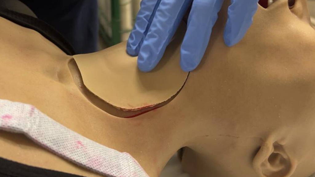
Researchers at the University of Minnesota Twin Cities have achieved a breakthrough in medical simulation: a new method for developing lifelike 3D-printed tissues that more accurately mimic the complexity of real human tissue. Where previous models were often rigid and simplistic, this innovation combines realistic strength, elasticity and tactile feedback. Properties that are essential for surgical training.
More realistic simulations
The researchers discovered how they could determine the mechanical properties of the printed material through controlled microstructures. An additional mathematical formula predicts how the tissue behaves under pressure or cutting movements. A technique was also developed to integrate blood-like fluids using microcapsules, which adds even more realism to simulations.
An initial evaluation shows that surgeons rate the new models significantly higher than conventional training materials, particularly because of the realistic resistance and response during cutting. This suggests that the technology can contribute to better training results and ultimately to safer patient care.
Future applications
The next step is to scale up the method and apply it to a wider range of organs. Think of bionic organs for training purposes or tissues that respond to surgical techniques such as electrocautery. This will create a new generation of training models that not only offer realism, but also respond to the diversity and complexity of real operations.
For medical training, this represents a significant step forward: surgeons and doctors in training will soon be able to practise in a more realistic, risk-free environment, without having to rely on limited cadaver models or less accurate simulations. The research, conducted in collaboration with the University of Washington, highlights how digital and biomedical technology are coming together to better prepare healthcare professionals for practice.
Technology for better training
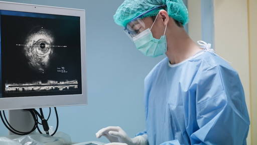
Earlier this year a research team at Mount Sinai, New York, developed an AI-driven training system that allows medical students to independently learn complex surgical procedures. The innovation combines deep learning algorithms with an extended reality (XR) headset, providing step-by-step visual instructions and real-time feedback directly to the trainee’s field of view. This approach addresses the shortage of qualified surgical trainers while reducing costs and ensuring more consistent teaching quality.
In a pilot study with 17 medical students, participants successfully performed a simulated partial nephrectomy on a 3D-printed “phantom kidney” filled with polymers to mimic human tissue. The AI analysed each action in real time, guiding the students with 99.9% accuracy during a critical step of the operation. According to lead investigator Dr. Nelson Stone, the study shows that autonomous AI-assisted surgical training is both feasible and effective, with the potential to make high-quality surgical education more accessible worldwide.


