Histology remains a cornerstone of medical diagnostics. By cutting tissue into ultrathin slices, staining it and examining it under a microscope, clinicians can determine whether tissue is diseased and guide treatment decisions. However, this classical approach is labour-intensive and inherently two-dimensional, disrupting the spatial structure of the tissue in the process.
Researchers are therefore exploring virtual histology: high-resolution, three-dimensional X-ray imaging that allows tissue to be examined intact. X-rays can penetrate samples several centimetres thick, enabling full-volume analysis and inspection from any angle. Yet until now, one major limitation remained: unlike conventional histology, X-ray images are monochrome, making it impossible to distinguish stained structures from surrounding tissue.
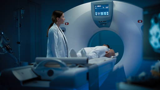
X-ray computed tomography
A new approach developed by Hereon Institute of Materials Physics and international partners offers a solution. Led by researcher Dominik John, the team combined high-resolution X-ray computed tomography with a phase-contrast technique and a novel analysis algorithm. Their work has been published in Advanced Science.
The method simultaneously measures two properties: how strongly tissue absorbs X-rays and how much it refracts them. Using a fine grid placed in the X-ray beam, the system generates two separate 3D datasets, one showing the tissue structure, the other isolating the dye. In effect, this introduces “colour” into X-ray imaging.
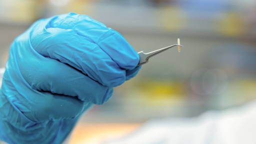
To demonstrate the technique, the researchers analysed kidneys from mice and rats stained with hematein, enhanced with a lead atom to improve X-ray contrast. Measurements were performed at the DESY PETRA III facility in Hamburg and the Australian Synchrotron in Melbourne. The system not only visualised where the dye was located, but also quantified its concentration across the tissue, closely matching results from conventional histology.
Support biomedical research
While the technique currently relies on large-scale research infrastructure, the team is working to adapt it for advanced laboratory X-ray sources. In the short term, the technology could support biomedical research, including oncology. In the longer term, further improvements in resolution could make it clinically relevant.
For healthcare, virtual histography could enable disease assessment in full spatial context, helping clinicians better evaluate tumour spread, surgical margins or treatment effects. Ultimately, this may lead to more precise diagnoses, better-informed therapies and less invasive procedures, offering clear benefits for both clinicians and patients.
3D-imaging
Last year, researchers from Politecnico di Milano developed the world’s first free online application that helps healthcare professionals select the most suitable AI model for generating high-quality 3D images of specific organs. Led by Dr. Andrea Moglia, the project represents an important step toward more efficient and precise medical imaging.
The tool allows clinicians to choose an organ or anatomical region and immediately see which validated AI models perform best for accurate image segmentation. This reduces trial and error, limits human bias and speeds up the creation of reliable 3D reconstructions from CT or radiographic data.