Philips announced the launch of DeviceGuide, an artificial intelligence (AI)-driven innovation designed to support physicians in treating a leaky mitral valve without open-heart surgery. By turning live imaging data into an intuitive 3D view inside the beating heart, DeviceGuide acts as an intelligent co-pilot for physicians, enhancing visibility, confidence, and precision during minimally invasive interventions.
AI sees what doctors couldn’t before
“For the first time, AI is assisting doctors in real time as they repair the human heart,” said Dr. Atul Gupta, Chief Medical Officer for Diagnosis & Treatment at Philips and practicing interventional physician.
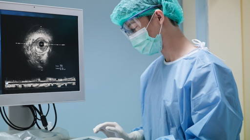
Repairing a leaky mitral valve, a condition known as mitral regurgitation, which affects an estimated 37 million adults worldwide, is one of medicine’s most technically demanding tasks. Traditionally, physicians have relied on flat, two-dimensional X-ray and ultrasound images, mentally reconstructing the patient’s anatomy as they guide catheters and implants through the moving chambers of the heart.
DeviceGuide redefines how this procedure is performed. By using deep-learning algorithms to interpret and fuse X-ray and ultrasound images in real time, it creates a live, 3D visualization of the heart and the device’s position within it.
“Imagine trying to hit a target inside a moving tennis ball, while it’s spinning and its walls are transparent,” Dr. Gupta explained. “That’s what guiding a catheter through a beating heart feels like. DeviceGuide changes that completely. It works like a kind of intelligent co-pilot, a system that tracks the device inside the heart, keeps it in focus, and shows you exactly where you are and where you need to go.”
Less invasive heart procedures in 3D
At the core of DeviceGuide is AI-based device intelligence that automatically recognizes, tracks, and highlights the implant’s silhouette as it moves through the heart. Every small movement is analyzed in real time, helping physicians navigate with precision and maintain orientation at all times. The AI also fuses multiple imaging views into a single, coherent 3D display, so that every member of the heart team, including the echocardiographer and the interventional cardiologist, sees the same, synchronized, real-time visualization.
“It’s as if a beacon of light were attached to the device itself: always visible, always in focus. The AI tracks its 3D movement through the heart’s chambers, showing its position and direction in real time. That gives doctors a clear sense of depth and orientation that wasn’t possible before,” according to Dr. Gupta.
DeviceGuide is designed for interventional cardiologists, structural heart specialists, echocardiographers, and cath-lab teams performing transcatheter mitral valve repair procedures, known as TEER (Transcatheter Edge-to-Edge Repair). The solution aims to simplify these interventions, supporting both highly experienced centers and hospitals newer to structural heart therapies.
By providing clear, consistent visual guidance, DeviceGuide helps reduce complexity and shortens the learning curve. Hospitals around the world, including those in developing regions or smaller healthcare systems, could potentially expand treatment access to patients who might otherwise go untreated due to a lack of experience or resources.
AI-enhanced Image-Guided Therapy
DeviceGuide builds on Philips’ long legacy in image-guided therapy (IGT), a field that enables physicians to perform complex procedures through small incisions using live imaging rather than open surgery. Every second, somewhere in the world, a patient is treated using Philips’ image-guided therapy technology.
DeviceGuide helps clinicians make precise decisions in the moment during live interventional procedures. This marks a major step for Philips, as artificial intelligence transitions from a diagnostic assistant in imaging or monitoring to a real-time decision-support tool during the procedure itself.
During a mitral valve repair, teamwork and timing are everything. One physician controls the imaging probe, another navigates the catheter, and others monitor live data. DeviceGuide creates one shared visual reference, showing everyone in the room the same orientation of the anatomy and device. This can streamline communication, reduce manual adjustments, and help the team stay focused on the patient rather than on imaging controls.

AI is actively helping to guide the procedure as it happens
The system runs on Philips’ Azurion image-guided therapy platform and its EchoNavigator real-time imaging fusion technology. Together, they combine live ultrasound and X-ray views to visualize both soft tissue and devices. DeviceGuide’s deep-learning engine continuously analyzes these inputs, tracking the implant’s position, trajectory, and orientation throughout the procedure, effectively functioning as a GPS for the heart.
DeviceGuide was developed in collaboration with Edwards Lifesciences, a global leader in structural heart innovations. The partnership combines Edwards’ expertise in heart valve therapies with Philips’ leadership in imaging and AI-based guidance systems. Together, the companies have optimized parts of the mitral TEER workflow, supporting heart teams with the latest clinical and technological insights.
For patients, minimally invasive procedures guided by AI may mean faster recovery, fewer complications, and access to life-saving treatment for those too frail for open-heart surgery. For clinicians, DeviceGuide could reduce cognitive load, improve accuracy, and foster teamwork through shared real-time visualization.
“This is AI built for the realities of the procedure room. It understands the rhythm of the heart, the motion of the device, and supports the entire team moment by moment.”


