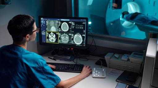
A routine retinal photograph taken during an eye exam may soon do more than assess vision. New research from the University of Florida suggests that artificial intelligence can use these images to identify a range of risk factors associated with Alzheimer’s disease, potentially offering a low-cost tool for earlier detection and prevention.
The study analyzed retinal images from more than 40,000 individuals and found that subtle changes in the retina can reveal biological and lifestyle factors linked to future dementia risk. One of the greatest challenges in Alzheimer’s disease is timing. By the time symptoms become apparent and conventional diagnostic tests confirm the disease, significant and often irreversible brain damage has already occurred.
Beyond traditional diagnostics
According to lead researcher Ruogu Fang, professor of biomedical engineering at the University of Florida, there is growing interest in identifying earlier biomarkers that can signal vulnerability long before cognitive decline becomes visible. “Alzheimer’s develops over decades,” Fang explained. “Retinal health may offer an opportunity to identify at-risk individuals much earlier and encourage preventive interventions, lifestyle changes and further testing.”
The retina, an extension of the central nervous system, shares important biological characteristics with the brain. As a result, changes in retinal structures may reflect broader neurological and vascular processes associated with neurodegenerative disease. The research was recently published in the Journal of Alzheimer’s Disease.
Unlike MRI scans or other advanced imaging techniques, retinal photographs are already widely used in routine clinical practice. Patients with diabetes, glaucoma, cataracts or other eye conditions often undergo retinal imaging regularly, while many optometry clinics also capture retinal photographs during standard eye examinations. This widespread availability makes retinal imaging an attractive candidate for large-scale screening. Compared with brain imaging technologies, retinal scans are inexpensive, non-invasive and easy to perform.
Using machine learning algorithms, researchers analyzed retinal photographs from a large UK-based patient database. The AI system identified specific retinal regions, including blood vessels and the optic nerve, that were associated with known Alzheimer’s risk factors. According to first author Seowung Leem, a doctoral researcher at the University of Florida, AI enables the detection of subtle retinal variations that would be difficult for clinicians to identify manually across such large populations.
Risk factors hidden in the eye
The AI model successfully predicted several biological characteristics and lifestyle-related risk factors known to influence Alzheimer’s disease risk. Among the factors accurately identified were sex, blood pressure, smoking behavior, alcohol consumption and insomnia. Many of these variables are typically collected through medical records or patient questionnaires, but such information can be incomplete or dependent on self-reporting.
Retinal imaging offers a more objective alternative. Importantly, retinal changes may also reflect the cumulative effects of long-term exposure to risk factors, providing a biological record that goes beyond a patient’s current health status. Fang describes the retina as an “integrated biological sensor” capable of capturing neurovascular health and cumulative disease vulnerability rather than merely serving as a substitute for lifestyle questionnaires.
Earlier intervention
The findings build on previous research from Fang’s team demonstrating that retinal images can help identify individuals who already have Alzheimer’s disease. The new study shifts the focus to an even earlier stage: detecting risk before symptoms emerge. Researchers believe this could have important implications as new Alzheimer’s therapies become available. Earlier identification of at-risk individuals could enable interventions ranging from lifestyle modifications and preventive treatments to cognitive training programs before significant brain damage occurs.
While further validation is needed before retinal AI screening can be incorporated into routine care, the study highlights the growing potential of combining ophthalmology, artificial intelligence and neurology. A simple eye scan may one day become a valuable tool for identifying those most likely to benefit from early Alzheimer’s prevention strategies.
Similar breakthrough
Last year, researchers at the National University of Singapore (NUS Medicine) achieved a similar breakthrough developing RetiPhenoAge, an AI-powered digital biomarker that uses a simple retinal photograph to predict the risk of cognitive decline and dementia. By applying deep learning to standard eye scans, the technology estimates the retina’s “biological age,” which is closely linked to brain health and ageing processes.
In a study involving more than 500 memory-clinic patients in Singapore, individuals with an older retinal age faced a 40% higher risk of cognitive decline within five years. The findings were further validated using data from over 33,000 participants in the UK Biobank followed for 12 years. A key advantage of RetiPhenoAge is its scalability and accessibility. Because retinal imaging equipment is already widely available in primary care and eye clinics, the technology offers a non-invasive, cost-effective screening tool.