For more than 150 years, pathology has relied on a familiar process: thin slices of tissue are stained with chemical dyes and examined under a microscope to identify disease. Now, researchers have demonstrated a new approach that could eventually transform this workflow. By combining high-resolution micro-computed tomography (micro-CT) with artificial intelligence, scientists have created three-dimensional virtual tissue stains that closely resemble conventional histology images.
The proof-of-concept study, led by researchers at the Paul Scherrer Institute (PSI) in Switzerland and published recently, suggests that future pathology workflows may no longer depend exclusively on fragile tissue sections and labor-intensive staining procedures.
Bringing color to 3D tissue imaging
Histology remains the gold standard for diagnosing many diseases, from cancer to cardiovascular disorders. The process, however, is inherently two-dimensional. Tissue samples must be sliced into ultrathin sections, stained with dyes and examined under a microscope. The PSI-led team sought to overcome this limitation by using phase-contrast micro-computed tomography (PCµCT), an advanced imaging technique capable of visualizing soft tissue structures in three dimensions at microscopic resolution.
While PCµCT provides highly detailed images, the resulting datasets are displayed in grayscale. This presents a challenge because pathologists are trained to interpret the characteristic color patterns produced by traditional stains. Cell nuclei, collagen fibers and other tissue structures are often identified through these familiar visual cues.
To bridge this gap, the researchers developed VISTACT (Virtual Staining of Micro-Computed Tomography), an AI-powered platform that translates grayscale CT images into colorized virtual histology images. “We have shown for the first time that a CT-based virtual stain can deliver results similar to conventional laboratory histology,” said physicist Goran Lovric, who led the study. “This could open up a wealth of clinical and scientific applications.”
Recognize tissue patterns
The system was trained using paired datasets consisting of conventional histological sections and matching CT scans. By comparing the two image types, the AI learned which microscopic structures typically correspond to specific staining patterns.
To achieve this, the team used a conditional generative adversarial network (cGAN), a machine-learning model designed for image-to-image translation. The AI generated detailed virtual stains that reproduced many of the visual characteristics pathologists rely on in routine practice.
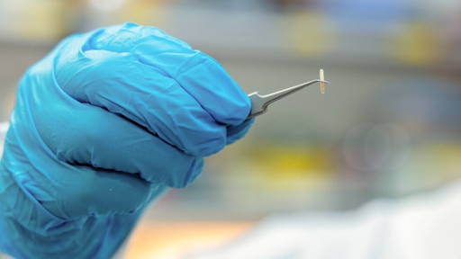
According to the researchers, the virtual images were able to distinguish a variety of tissue components, including collagen structures, blood vessels and lung surfaces. Achieving this required highly precise alignment between physical tissue sections and their corresponding locations within the three-dimensional CT volume. The team developed a multi-step image registration process to accurately map each microscopic section back into the CT dataset, improving on existing alignment methods.
Potential research applications
To evaluate the technology, the researchers applied VISTACT to lung tissue samples from patients with pulmonary hypertension, a condition characterized by structural changes in pulmonary blood vessels. The virtual staining technique enabled researchers to visualize diseased vascular regions in three dimensions, offering perspectives that are difficult to obtain using conventional histology alone.
Because the method preserves the tissue sample intact, it may prove particularly valuable for studying tumors, vascular disorders and other diseases where understanding complex three-dimensional tissue architecture is important. Researchers believe the technology could eventually accelerate biomarker discovery and support the development of new diagnostic approaches by providing a more comprehensive view of tissue organization.
Early stage
Despite the encouraging results, the researchers emphasize that the technology remains at an early stage. The imaging was performed using the TOMCAT beamline at the Swiss Light Source, a specialized research facility not available in routine clinical practice. In addition, the generated images remain statistical predictions rather than direct measurements. While the AI can produce highly plausible virtual stains, it does not create actual histological information.
Resolution limitations also remain. Current datasets are not always detailed enough to reliably visualize individual cell nuclei, an essential requirement for many diagnostic applications. Nevertheless, the researchers consider the study an important milestone. By demonstrating that AI can translate three-dimensional CT data into familiar histological representations, they have established a foundation for future developments in digital pathology. More than a century after Rudolf Virchow's pioneering work helped establish modern pathology, advances in imaging and artificial intelligence may be paving the way for a new era of non-destructive, three-dimensional tissue analysis.
Earlier this year we reported another approach, developed by Hereon Institute of Materials Physics and international partners. Researchers combined high-resolution X-ray computed tomography with a phase-contrast technique and a novel analysis algorithm.