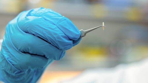
A newly developed 4D heart model is showing promise in improving treatment outcomes for patients with heart failure undergoing cardiac resynchronization therapy (CRT). The technology, developed at the University of Calgary, was evaluated in a clinical trial.
The model uses cardiac MRI data to create a patient-specific digital twin of the heart. These dynamic, “beating” 3D representations allow clinicians to better guide the placement of CRT pacemakers, which are used to restore coordinated contraction of the heart muscle in patients with impaired cardiac function. The clinical trial was published in Circulation: Arrhythmia and Electrophysiology.
Personalised guidance for CRT
CRT is a well-established therapy for advanced heart failure, but up to one-third of patients do not respond adequately to standard implantation techniques. The MAPIT-CRT trial investigated whether using a personalised digital heart model could improve treatment effectiveness.
In the study, 202 patients across seven Canadian centres were followed for six months after receiving CRT. Patients whose treatment was guided by the 4D model showed significantly better outcomes compared to those receiving standard care. Heart function improved by 10.8% in the model-guided group, compared to 5.8% in the control group. Additionally, 66% of patients in the intervention group experienced clinical improvement, versus 52% in the standard care group. Importantly, the use of the digital model did not increase procedure time, complication rates or recovery risks.
Combining imaging and computational modelling
According to the researchers, the study demonstrates how advanced imaging and computational modelling can be integrated into clinical practice to support more precise, patient-specific care. By simulating how different pacemaker placements affect heart function, clinicians can make more informed decisions during the procedure. Beyond CRT, the technology may have broader applications in cardiology, including earlier disease detection and improved prediction of patient outcomes.
A key focus in the development of the model was usability. Unlike earlier approaches that required complex software integration, the new system is designed as an accessible, web-based platform. This could facilitate wider adoption in clinical settings and accelerate the implementation of precision medicine in cardiology. The researchers conclude that digital twin technology represents a significant step towards more personalised and effective treatment strategies for heart failure patients.
3D heart model for surgical training
A couple of weeks ago we reported on the development of a 3D-printed heart model that can beat and contract, offering a realistic platform for surgical training. The fully synthetic model replicates the left side of the human heart, including key structures such as the atrium, ventricle and mitral valve, and mimics both anatomical form and dynamic function.
Using pneumatic actuators and artificial blood flow, the model responds to procedures in real time. In tests, researchers successfully performed a valve repair, validated by sensors and imaging. Unlike traditional training methods that rely on animal models or cadavers, the reusable model enables more flexible and ethical training. Researchers say the technology could improve training for complex and minimally invasive procedures, while providing a more accurate and patient-relevant simulation of cardiac function.